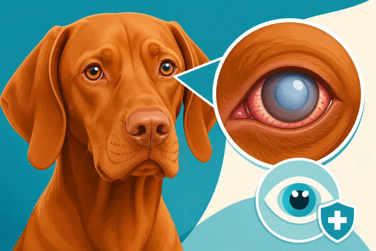
Eye Disease in a Vizsla
It’s a truly gut-wrenching moment for any Vizsla owner when you notice something isn’t quite right with their eyes. Our copper companions are known for their vibrant energy and keen focus, especially when working in the field or simply gazing up at you with those soulful, intelligent eyes. So, when they start squinting, bumping into furniture, or their eyes just look… off, it can feel like a punch to the gut. This comprehensive guide is here to walk you through the various eye diseases that can affect Vizslas, helping you understand the symptoms, diagnosis, and management options so you can best advocate for your beloved dog’s vision and comfort.
The Unique Vulnerability of Vizsla Eyes
Vizslas are a truly special breed, renowned for their intelligence, athleticism, and almost unparalleled devotion to their human families. But like many purebred dogs, their unique genetic makeup can unfortunately predispose them to certain health conditions, and eye diseases are certainly on that list. Understanding why they might be more vulnerable is the first step in being a proactive and informed owner.
Why Vizslas Might Be Prone to Eye Issues
The story of the Vizsla is one of survival and rebirth, having faced extinction multiple times. This dramatic bottleneck in their population, particularly during the two World Wars and under communist rule, meant that the breed was rebuilt from a very small gene pool. While dedicated breeders worked tirelessly to save the Vizsla, this reduced genetic diversity has inadvertently concentrated certain genes, making them more susceptible to specific health conditions, including those affecting their eyes. It’s not a fault of the breed, but a historical legacy that owners need to be aware of. We cherish these dogs for their history and their character, and that often comes with a responsibility to understand their potential health challenges.
Beyond genetics, a Vizsla's lifestyle naturally exposes them to more environmental hazards. These are not couch potatoes; they are active, athletic, and often spend hours running through fields, dense cover, and woods, pursuing game. This means their eyes are frequently exposed to dust, pollen, sharp twigs, burrs, and other debris that can cause irritation, scratches, or even more serious trauma. A simple day in the field can turn into an emergency vet visit if a foreign body lodges in the eye or causes a significant corneal abrasion. Their short, fine coats also offer less protection around the face compared to breeds with longer facial hair, leaving their eyes more exposed to the elements. So, while their heritage and drive are incredible, they do come with a practical need for vigilance regarding eye health.
Early Warning Signs Not to Miss
As a "Velcro Vizsla" owner, you're probably already intimately familiar with your dog's every sigh and twitch. This close bond is actually a huge advantage when it comes to spotting subtle changes in their health. Your Vizsla probably follows you into every room, probably even into the bathroom! This constant proximity means you're uniquely positioned to catch early warning signs of eye trouble that might go unnoticed in a less clingy breed.
One of the most concerning, yet sometimes overlooked, signs is a change in their behavior, especially their confidence and spatial awareness. If your usually fearless Vizsla starts hesitating on stairs, bumping into familiar furniture in dimly lit rooms, or seems disoriented when in a new environment, it could be an indicator of declining vision, particularly night vision. They might also become more reliant on you, sticking even closer than usual, as a way to navigate their world when their sight is compromised. Don’t dismiss these as just "being clumsy" or "getting old" without ruling out an underlying eye issue.
Beyond behavioral cues, there are physical changes in the eye itself that warrant immediate attention. Look for any redness or inflammation of the white part of the eye (sclera) or the pink lining of the eyelids (conjunctiva). Excessive tearing or, conversely, very dry-looking eyes with thick, sticky discharge are also red flags. Squinting, holding one eye partially or completely closed, or rubbing at their face can indicate pain or irritation. Any cloudiness, discoloration, or change in the size or shape of the pupil (the black center) should also prompt a vet visit. Remember, early intervention can often make a significant difference in the outcome of many eye conditions, so trusting your gut and getting it checked out by a veterinarian or a veterinary ophthalmologist is always the best course of action.
Common Eyelid and Eyelash Conditions
The eyelids and eyelashes act as the first line of defense for a Vizsla's precious eyes. When something goes wrong with them, it can lead to constant irritation, pain, and even serious damage to the delicate surface of the eye. These conditions are often uncomfortable and can severely impact your dog’s quality of life if left untreated.
Entropion and Ectropion: Inward and Outward Turning Eyelids
Entropion is a condition where a portion of the eyelid, usually the lower one, rolls inward towards the eye. Imagine having an eyelash constantly rubbing against your eyeball – that's what your Vizsla experiences. The fur and lashes on the eyelid literally scrape against the cornea, leading to irritation, excessive tearing, and potentially painful corneal ulcers or scarring. This can be incredibly uncomfortable and can make your otherwise happy-go-lucky Vizsla miserable. While some dogs are born with this genetic predisposition, it can sometimes develop secondary to trauma, severe conjunctivitis, or even chronic squinting from other painful eye conditions. Vizslas, with their somewhat loose skin and expressive faces, can unfortunately be prone to this. You can learn more about this specific issue in our detailed article, Whether a Vizsla Gets Entropion.
Conversely, ectropion is when the eyelid rolls outward, exposing the delicate conjunctiva. This gives the eye a droopy appearance, sometimes making it look perpetually sad or tired. While it might not cause the same painful abrasion as entropion, ectropion leaves the eye vulnerable to dryness, irritation from environmental debris like dust and wind, and chronic infections because the eyelid can't properly protect the eye or distribute tears. The exposed conjunctiva often becomes red, thickened, and inflamed. Vizslas are not as commonly affected by severe ectropion as some other breeds (like Basset Hounds or Bloodhounds), but milder forms can certainly occur and contribute to chronic eye issues. If you notice a perpetually droopy or irritated eye, it's worth discussing with your vet. Our article, Whether a Vizsla Gets Ectropion, delves deeper into what this might mean for your dog.
Both entropion and ectropion are typically diagnosed through a thorough ophthalmic examination by your veterinarian. They’ll assess the structure of the eyelids, look for signs of irritation or corneal damage, and may use special stains to highlight any ulcers on the eye's surface. The primary treatment for both conditions, especially if they are causing significant discomfort or damage, is surgical correction. This involves a delicate procedure to remove a small wedge of tissue from the eyelid, effectively tightening or repositioning it to its correct alignment. While surgery might sound daunting, it's often a highly effective solution that provides immense relief and prevents further damage, allowing your Vizsla to return to a comfortable, active life without constant eye irritation.
Distichiasis and Ectopic Cilia: Misplaced Eyelashes
Beyond the eyelid structure itself, sometimes the eyelashes decide to grow in the wrong place, causing chronic irritation. Distichiasis is a condition where extra eyelashes (distichia) grow from abnormal locations on the eyelid margin, often from the ducts of the meibomian glands, which are usually responsible for producing the oily component of tears. These extra lashes can range from soft and harmless to stiff and abrasive, constantly rubbing against the surface of the eye (the cornea) and causing chronic irritation, tearing, and even corneal ulcers. It’s like having a tiny, sharp piece of hair poking your eye every time you blink. Vizslas, like many breeds, are genetically predisposed to this condition, and it often appears when they are young.
Ectopic cilia are even more problematic. These are single, usually stiff, eyelashes that grow directly through the conjunctiva (the pink lining of the eyelid) and protrude onto the surface of the eye. Because they grow straight out and are often quite rigid, they cause intense pain and irritation, almost always leading to corneal ulcers. These are extremely painful and can develop very quickly, often requiring immediate veterinary attention. Your Vizsla might present with severe squinting, profuse tearing, and pawing at the affected eye. It’s a bit like having a thorn embedded in your eyelid, constantly pricking your eye.
Diagnosis of both conditions typically involves a detailed examination of the eyelid margins, often using magnification, to pinpoint the offending lashes. For ectopic cilia, the lash can be surprisingly hard to spot without the right equipment and experience, as it often hides just under the eyelid. Treatment aims to permanently remove the abnormal lashes. For distichiasis, options include cryotherapy (freezing the hair follicles), electrocautery (burning the follicles), or surgical removal of the affected follicle. Ectopic cilia are usually treated by surgically excising the follicle through the conjunctiva to prevent regrowth. While seemingly minor, these misplaced lashes can cause significant discomfort and damage, and addressing them properly can bring enormous relief to your Vizsla.
Conditions Affecting the Cornea and Lens
The cornea, the clear outer window of the eye, and the lens, responsible for focusing light, are critical components for clear vision. When these structures are compromised, it can lead to anything from mild cloudiness to severe vision loss.
Corneal Dystrophy: Cloudiness and Vision Impairment
Corneal dystrophy refers to a group of inherited, non-inflammatory conditions that cause deposits of abnormal material (usually lipids or minerals) within one or more layers of the cornea. Essentially, the clear window of your Vizsla's eye can start to look cloudy or hazy. This isn't usually a painful condition in itself, but it can certainly affect vision, especially if the deposits are dense or widespread. There are different types: epithelial, stromal, and endothelial, named after the layer of the cornea where the deposits occur. Stromal corneal dystrophy, for instance, often appears as a milky white or crystalline opacity in the middle layer of the cornea.
Symptoms can vary based on the type and severity. You might simply notice a subtle cloudiness or a dull, non-reflective patch on the surface of your Vizsla's eye that doesn't seem to bother them. In more advanced cases, however, these opacities can significantly impair vision, making it difficult for your dog to navigate, especially in low light. Some forms of epithelial dystrophy can lead to recurrent corneal erosions, which are painful, causing squinting and tearing, and can predispose the eye to secondary infections. It’s important not to confuse corneal dystrophy with cataracts, which affect the lens, or corneal edema, which is usually a sign of an underlying issue.
Diagnosing corneal dystrophy involves a thorough ophthalmic examination by a vet or veterinary ophthalmologist. They will use a specialized light source and magnification (like a slit lamp) to examine the corneal layers and characterize the deposits. Since it's an inherited condition, genetic testing might become available for certain forms in the future for Vizslas, though specific tests for all types aren't always routine yet. Treatment for corneal dystrophy is often supportive rather than curative, as the underlying cause is genetic. For dogs with vision impairment due to dense deposits, a superficial keratectomy (surgical removal of the outer corneal layers) can sometimes improve clarity, but the deposits may recur. Managing any secondary corneal erosions with lubricating drops or ointments is also crucial for comfort.
Cataracts: Clouding of the Lens
Cataracts are perhaps one of the most well-known eye conditions, affecting the lens of the eye. A normal lens is perfectly clear, allowing light to pass through and focus on the retina. A cataract is any opacity or clouding within the lens, which then obstructs light and causes vision to become blurry or, in severe cases, completely lost. It’s like looking through a frosted window. This is a common cause of vision impairment in older dogs, but it’s important to understand that cataracts aren't always just an "old age" thing. You can learn more about this in our article Whether a Vizsla Gets Cataracts.
Symptoms of cataracts are fairly straightforward: you’ll notice a milky, cloudy, or bluish-white appearance within the pupil (the center of the eye). This opacity will be behind the clear surface of the cornea. As the cataract progresses, your Vizsla's vision will decline. They might start bumping into objects, especially in unfamiliar surroundings or at dusk. Their confidence in navigating known spaces might decrease, and they might rely more on their other senses. Sometimes, a Vizsla might not show obvious signs until their cataracts are quite advanced because they compensate so well and their other senses are so acute.
Cataracts can arise for several reasons in Vizslas. Senile cataracts, common in older dogs, develop simply due to aging. However, Vizslas can also develop inherited or juvenile cataracts, appearing in younger dogs, sometimes even as puppies. These often have a genetic basis, making careful breeding practices and health clearances important. Cataracts can also be secondary to other diseases, most notably diabetes mellitus (often called diabetic cataracts), or as a result of trauma to the eye or certain inflammatory conditions. Diagnosis is made through a comprehensive ophthalmic examination, often including dilation of the pupils to get a clear view of the lens. For Vizslas with cataracts significantly affecting their vision, surgical removal of the cataract (phacoemulsification, similar to human cataract surgery) can restore sight. This is a specialized procedure with a high success rate, but it requires careful patient selection and post-operative care.
Lens Luxation: Dislocated Lenses
Lens luxation is a serious and often painful condition where the lens of the eye becomes partially or completely dislocated from its normal position. The lens is held in place by tiny fibrous structures called zonules. When these zonules break down or tear, the lens can move – either partially (subluxation) or completely (luxation) – into the anterior chamber (front of the eye) or posterior chamber (back of the eye). This is an emergency situation, as it can quickly lead to severe pain, inflammation, and secondary glaucoma.
There are two main categories of lens luxation: primary and secondary. Primary lens luxation is often inherited and due to a congenital weakness or degeneration of the zonular ligaments. While terriers are famously predisposed, Vizslas can also be affected, making it crucial for breeders to be aware of any family history of this condition. Secondary lens luxation, on the other hand, occurs as a result of trauma, chronic glaucoma, or severe inflammation within the eye that damages the zonules. For an active Vizsla, a forceful impact or severe eye injury could potentially lead to this.
Symptoms of lens luxation are typically acute and dramatic. Your Vizsla will likely show sudden, intense pain in the affected eye, manifesting as severe squinting, profuse tearing, and pawing at their face. The eye itself will usually be very red, and the cornea may appear cloudy or hazy. If the lens has moved into the anterior chamber, you might even be able to see it sitting abnormally in front of the iris. Vision will be severely compromised or completely lost in the affected eye. This is not a wait-and-see condition; it requires immediate veterinary attention. Diagnosis involves a thorough ophthalmic exam, often with specialized instruments. Treatment is usually surgical, involving the removal of the luxated lens to alleviate pain and prevent secondary complications like glaucoma. Without prompt intervention, a luxated lens can lead to permanent blindness and even necessitate eye removal due to uncontrollable pain.
Retina and Optic Nerve Diseases
The retina and optic nerve are the "film" and "cable" of the eye, respectively, responsible for capturing light and sending those images to the brain. Diseases affecting these crucial structures often lead to irreversible vision loss.
Progressive Retinal Atrophy PRA: Gradual Blindness
Progressive Retinal Atrophy, or PRA, is a group of inherited degenerative diseases that affect the photoreceptor cells (rods and cones) in the retina. Simply put, the cells that detect light gradually die off, leading to irreversible blindness. It's a heartbreaking diagnosis because it inevitably leads to complete vision loss, often over months or a few years. For an active, intelligent breed like the Vizsla, losing their sight can be incredibly challenging, both for them and their dedicated owners. You can read more about this in our specific article, Whether a Vizsla Goes Blind From PRA.
The typical progression of PRA starts with night blindness. The rod cells, responsible for vision in low light, are usually affected first. You might notice your Vizsla becoming hesitant to go out in the dark, bumping into things at night, or struggling to find their toys in dimly lit rooms. As the disease advances, cone cells (responsible for day vision and color perception) also degenerate, leading to daytime blindness. Other symptoms include increasingly dilated pupils, which may appear 'shiny' in low light (due to a reflective layer behind the retina called the tapetum lucidum), and sometimes secondary cataracts develop as a complication. It’s a slow, insidious process that can be difficult for owners to come to terms with.
Diagnosis of PRA is primarily made through a specialized test called an electroretinogram (ERG), which measures the electrical activity of the retinal cells in response to light. This can detect the disease even before clinical signs are apparent. Genetic testing is available for specific forms of PRA in many breeds, and while specific tests for all forms in Vizslas may still be developing, it's an important tool for breeders to screen their dogs and prevent passing on the disease. Unfortunately, there is no cure or effective treatment for PRA; the degeneration is irreversible. Management focuses on helping your Vizsla adapt to their progressive vision loss by maintaining a consistent home environment, using verbal cues, and ensuring their safety. It requires a tremendous amount of patience and love, but Vizslas are incredibly resilient and can adapt remarkably well with the right support.
Optic Nerve Hypoplasia: Underdeveloped Optic Nerves
The optic nerve acts as the vital communication cable between the eye and the brain. Optic nerve hypoplasia is a congenital condition, meaning it’s present from birth, where the optic nerve is underdeveloped or abnormally small. Instead of a robust bundle of nerve fibers, there are fewer or sometimes no functional fibers to transmit visual information. This can affect one eye (unilateral) or both eyes (bilateral) and results in partial or complete blindness in the affected eye(s).
The symptoms of optic nerve hypoplasia are evident from puppyhood. If bilateral, the puppy will be blind from birth, often showing signs of disorientation, bumping into objects, and an inability to track toys or people visually. Their pupils may appear dilated and unresponsive to light. If only one eye is affected, the other eye might compensate, making it harder to spot in a young, boisterous Vizsla puppy until one eye is specifically tested or the puppy encounters an obstacle it can't navigate with its good eye. This can be particularly frustrating for owners as they watch their puppy struggle with the world, unable to understand why.
Diagnosis involves a careful ophthalmoscopic examination by a veterinary ophthalmologist. They will observe the optic disc (the head of the optic nerve at the back of the eye) and note its small size or abnormal appearance. An ERG might also be performed to assess retinal function, which is usually normal, confirming the issue lies with the nerve itself. Unfortunately, there is no treatment for optic nerve hypoplasia, as the nerve tissue cannot be regenerated or repaired. For dogs with unilateral hypoplasia, their quality of life can be excellent as they adapt to relying on their one good eye. For those with bilateral blindness, adapting the home environment and relying heavily on other senses, as with PRA, becomes paramount. Understanding this condition early can help prepare owners for the unique challenges and joys of raising a blind Vizsla.
Glaucoma and Dry Eye
These two conditions represent opposite ends of the spectrum when it comes to fluid balance in the eye – one involves too much pressure, the other too little moisture. Both, however, can cause significant pain and vision problems for your Vizsla.
Glaucoma: Pressure Build-Up in the Eye
Glaucoma is an incredibly painful and serious eye condition characterized by abnormally high pressure inside the eye (intraocular pressure, IOP). Imagine a balloon being constantly over-inflated; that’s what happens to the eye. This increased pressure damages the optic nerve and retinal cells, leading to rapid and irreversible blindness if not treated promptly. It’s an emergency that demands immediate veterinary attention, as every hour counts in trying to save vision.
Glaucoma can be primary or secondary. Primary glaucoma is inherited and results from a structural defect in the eye's drainage angle, preventing fluid (aqueous humor) from leaving the eye properly. Certain breeds are genetically predisposed, and while Vizslas are not as commonly listed as primary glaucoma breeds like Beagles or Basset Hounds, any breed can theoretically be affected. Secondary glaucoma is much more common and occurs due to another underlying eye condition that blocks the drainage angle, such as a dislocated lens (lens luxation), severe inflammation (uveitis), or intraocular tumors. Given a Vizsla's active nature, an untreated eye injury could potentially lead to secondary glaucoma.
The symptoms of acute glaucoma are often dramatic and distressing. Your Vizsla will typically show severe pain, often crying out or pawing at the affected eye, which will likely be very red and squinted. The cornea may appear cloudy or bluish (corneal edema) due to the pressure. The pupil might be dilated and unresponsive to light. The eyeball itself might feel hard to the touch or even appear visibly enlarged in chronic cases. This is not a subtle condition; it’s an emergency. Diagnosis requires an immediate measurement of the intraocular pressure using a tonometer. Treatment is geared towards rapidly reducing the eye pressure with medications (drops and oral) to alleviate pain and save vision. In some cases, surgical procedures may be performed to improve drainage or reduce fluid production. Unfortunately, even with aggressive treatment, many dogs eventually lose vision in the affected eye due to the sensitive nature of the optic nerve to pressure damage. Early recognition and veterinary intervention are absolutely critical for any chance of retaining vision.
Keratoconjunctivitis Sicca KCS or Dry Eye
Keratoconjunctivitis Sicca, mercifully shortened to KCS or simply "dry eye," is a common condition where the eye doesn't produce enough tears. Tears are vital for lubricating the eye, flushing away debris, and providing oxygen and nutrients to the cornea. Without adequate tears, the eye becomes chronically dry, irritated, and susceptible to infection and corneal damage. It's a bit like driving a car without engine oil – things quickly start to seize up and get damaged. You can dive deeper into this topic with our article, Whether a Vizsla Gets Dry Eye.
Symptoms of KCS typically include chronic redness of the conjunctiva, frequent blinking or squinting, and a thick, yellowish-green discharge that can sometimes resemble pus. This discharge often accumulates in the corner of the eye and can be difficult to wipe away. Over time, the cornea might appear dull or hazy, and in chronic cases, pigment can spread across the cornea, leading to vision impairment. Vizslas with KCS will often rub their eyes due to the constant irritation. They might also develop recurrent bacterial eye infections because the natural antibacterial properties of tears are absent. This is a very uncomfortable condition that can significantly diminish a dog's quality of life, making every blink an irritating experience.
Diagnosing KCS is straightforward using a Schirmer Tear Test. A small strip of filter paper is placed under the lower eyelid for one minute, and the amount of moisture absorbed indicates the tear production. Low readings confirm KCS. The cause of KCS is often immune-mediated, meaning the body's own immune system attacks the tear glands. While it can also be caused by certain medications, nerve damage, or other diseases, the immune-mediated form is very common in many breeds, and Vizslas can be affected. Treatment for KCS is typically lifelong and involves medications that help stimulate tear production, most commonly cyclosporine or tacrolimus eye drops. These drops suppress the immune attack on the tear glands and encourage them to produce tears again. Artificial tears and lubricating ointments are also often used to provide immediate relief and comfort. Regular follow-up appointments are necessary to monitor tear production and adjust medication as needed. With consistent treatment, most dogs with KCS can live comfortably and avoid severe complications.
What to Expect at the Veterinary Ophthalmologist
When your Vizsla has an eye problem, your regular veterinarian is always the first point of contact. However, for complex, chronic, or vision-threatening conditions, they may recommend a consultation with a board-certified veterinary ophthalmologist. These specialists have advanced training and equipment specifically for diagnosing and treating eye diseases, and they can offer the most accurate diagnosis and advanced treatment options.
The Comprehensive Eye Exam Process
A visit to a veterinary ophthalmologist is a detailed experience, far more in-depth than a quick look at your Vizsla's eyes during a routine checkup. The process usually begins with a thorough discussion of your dog's history. Be prepared to share everything: when you first noticed symptoms, how they've progressed, any prior treatments, and their general health and medication history. This initial conversation is crucial for the ophthalmologist to start piecing together the puzzle.
Next comes the hands-on examination, which typically involves a series of specialized tests. The Schirmer Tear Test, which we discussed for KCS, is often one of the first, measuring tear production. Then, tonometry will be performed to measure the intraocular pressure, checking for glaucoma. The ophthalmologist will then use various tools to examine the different structures of the eye. A direct and indirect ophthalmoscope allows them to view the retina, optic nerve, and vitreous humor at the back of the eye. A slit lamp biomicroscope is particularly important; this instrument provides magnified, illuminated views of the eyelids, conjunctiva, cornea, iris, and lens, allowing for the detection of subtle abnormalities that would be impossible to see with the naked eye. Fluorescein staining, where a special orange dye is applied to the eye, is also routine to highlight any corneal abrasions or ulcers, as the dye sticks to damaged areas. It’s a comprehensive, systematic approach to ensure no detail is missed.
Understanding Diagnostic Tools and Genetic Testing
Beyond the standard eye exam, veterinary ophthalmologists have access to even more advanced diagnostic tools. If a retinal disease like Progressive Retinal Atrophy (PRA) is suspected, an electroretinogram (ERG) may be recommended. This test, often performed under light sedation, measures the electrical responses of the retina to light, providing a definitive diagnosis of retinal function. Ocular ultrasound is another valuable tool, especially if the internal structures of the eye (like the retina or lens) cannot be clearly visualized due to severe corneal opacity or hemorrhage. It can detect retinal detachments, tumors, or lens luxations hidden from direct view.
Genetic testing is becoming increasingly important, especially for breed-specific conditions. For Vizslas, understanding the genetic predispositions for conditions like PRA or certain types of cataracts can be vital, not just for the affected dog but also for breeding programs. Responsible breeders use genetic tests to screen their breeding stock, aiming to reduce the incidence of inherited diseases in future generations. When considering a Vizsla puppy, it's always wise to ask about the health clearances of the parents, including eye certifications. Organizations like the Orthopedic Foundation for Animals (OFA) maintain a public database of health certifications, including eye exams performed by certified veterinary ophthalmologists. These results contribute to a Canine Health Information Center (CHIC) number, indicating comprehensive health screening. You can learn more about these preventative measures in our article, The Health Tests Both Vizsla Parents Should Have, and further explore the value of a CHIC number in Whether a Vizsla Breeder Should Show a CHIC Number. These proactive steps by breeders are invaluable in safeguarding the breed's health.
Living with a Vizsla with Eye Disease
Discovering your Vizsla has an eye disease, especially one that impacts their vision or requires ongoing management, can be overwhelming. But with patience, adjustments, and consistent care, your Vizsla can continue to live a fulfilling and happy life. They are remarkably resilient, and their deep bond with you will be their greatest asset.
Managing Daily Life with Vision Impairment
If your Vizsla is facing partial or complete vision loss, adapting their daily environment is paramount to their safety and confidence. The number one rule: do not move furniture. Dogs with vision impairment create detailed mental maps of their homes using their other senses. Moving objects, even slightly, can cause confusion, anxiety, and lead to bumps and injuries. If you must rearrange, do it slowly and guide your Vizsla through the new layout repeatedly. Think about creating pathways with soft rugs or runners to help them navigate. Baby gates can be invaluable for blocking off stairs or unsafe areas, and ensuring dangerous objects are put away.
Outdoor spaces need careful consideration too. A fenced yard is ideal, ensuring no hazards like sharp objects or open water. When on walks, always keep your vision-impaired Vizsla on a leash. This isn't just for their safety from traffic or other animals, but also to prevent them from walking into obstacles they can't see. Consistent verbal cues become incredibly important: "step up," "step down," "wait," "danger," "this way." Your voice becomes their guiding light. Many owners find it helpful to introduce distinct scents in key areas (e.g., vanilla extract on furniture legs) to help their dog orient themselves. Vizslas are already so attuned to their owners that these adaptations often come quite naturally to them, and they respond beautifully to your calm, reassuring voice. The goal is to build their confidence, not foster dependency. Even if they are still hunting, an old Vizsla that still wants to hunt will need extra considerations for safety if their eyesight is failing.
Financial Considerations and Insurance
Let's be realistic: veterinary care for chronic or surgical eye conditions can be expensive. A consultation with a veterinary ophthalmologist alone can be hundreds of dollars, and specialized procedures like cataract surgery or glaucoma management can easily run into the thousands. Ongoing medications for conditions like KCS can also add up over the years. It's a significant financial commitment, but one we willingly make for our furry family members.
To help manage these costs, many Vizsla owners consider pet insurance. Plans vary, but a good comprehensive policy can cover a substantial portion of emergency care, specialist visits, diagnostics, and even ongoing medication for conditions that aren't pre-existing. For instance, a provider like Healthy Paws offers extensive coverage that can significantly ease the financial burden of unexpected health issues, making advanced treatments more accessible. It’s definitely worth researching different providers and their policies carefully, paying close attention to deductibles, reimbursement rates, and any exclusions for pre-existing conditions. Starting a policy when your Vizsla is young and healthy can be a smart move, ensuring a wider range of coverage as they age.
Even with insurance, it’s always a good idea to have an emergency fund specifically for your Vizsla's healthcare. Unfortunately, Vizslas can be prone to several immune-mediated diseases, which can also be costly to manage, so a robust insurance policy or savings plan is a wise investment. You can explore this further in Insuring a Vizsla Against Immune Disease and get more insight into whether it's the right choice for you in Whether a Vizsla Is Worth Insuring at All. The peace of mind that comes from knowing you can provide the best possible care without devastating your savings is truly invaluable.
Vizsla Eye Conditions: At a Glance
Here’s a quick overview of some common eye conditions that can affect Vizslas, their symptoms, and general approaches to treatment. This table is for informational purposes and should not replace a veterinary diagnosis.
| Condition | Description | Common Symptoms | Typical Treatment |
|---|---|---|---|
| Entropion | Eyelid rolls inward, causing lashes/fur to rub cornea. | Squinting, excessive tearing, redness, corneal ulcers, chronic irritation. | Surgical correction (eyelid tuck). |
| Ectropion | Eyelid rolls outward, exposing conjunctiva. | Droopy eyelid, chronic redness, discharge, dry eye, susceptibility to foreign bodies. | Surgical correction (if severe), lubricating drops/ointments. |
| Distichiasis/Ectopic Cilia | Extra eyelashes growing abnormally, irritating cornea. | Tearing, squinting, redness, corneal ulcers, rubbing at eye, acute pain (ectopic cilia). | Cryotherapy, electrocautery, surgical removal of follicles. |
| Corneal Dystrophy | Inherited, non-inflammatory deposits in cornea, causing cloudiness. | Hazy or cloudy patch on cornea, vision impairment (variable), sometimes recurrent erosions. | Often no treatment needed; keratectomy for severe vision loss; lubricants for erosions. |
| Cataracts | Clouding of the eye's lens. | Milky/bluish-white opacity in pupil, reduced vision, bumping into objects. | Surgical removal (phacoemulsification) to restore vision. |
| Lens Luxation | Lens dislocates from its normal position. | Sudden, severe pain, redness, squinting, cloudy cornea, vision loss, abnormal lens position. | Emergency surgical removal or repositioning of the lens. |
| Progressive Retinal Atrophy | Inherited degeneration of retinal cells, leading to blindness. | Night blindness progressing to daytime blindness, dilated pupils, often reflective eyes in dark. | No cure; management focuses on environmental adaptation. |
| Optic Nerve Hypoplasia | Underdeveloped optic nerve from birth. | Blindness (partial or complete) from puppyhood, dilated pupils, unresponsive to light. | No treatment; management focuses on environmental adaptation. |
| Glaucoma | High pressure inside the eye, damaging optic nerve. | Severe pain, redness, squinting, cloudy cornea, dilated pupil, enlarged eyeball (chronic), rapid vision loss. | Emergency medical drops/oral meds to reduce pressure, sometimes surgery. |
| Keratoconjunctivitis Sicca | Insufficient tear production (dry eye). | Chronic redness, thick discharge, squinting, corneal changes (pigment/vascularization), recurrent infections. | Lifelong tear-stimulating medications (e.g., cyclosporine), artificial tears. |
Disclaimer
This article is intended for informational purposes only and does not constitute veterinary advice. If you suspect your Vizsla is experiencing any eye issues, please consult with a qualified veterinarian or a board-certified veterinary ophthalmologist immediately. Early diagnosis and treatment are crucial for the best possible outcome for your dog's vision and comfort.